Introduction to Diagnostic Medical Parasitology
Wuchereria bancrofti
Microfilaria of Wuchereria bancrofti

The microfilariae of Wuchereria bancrofti have several distinguishing characteristics. They are sheathed and their size varies depending on the medium used for staining: measuring 240—300 µm in stained blood smears and 275—320 µm in 2% formalin.
The body of these microfilariae is gently curved, and their tails taper to a point. Within the microfilaria's body, the nuclear column, which consists of the cells that make up the microfilaria, is loosely packed. As a result, individual cells can be visualized separately, and they do not extend all the way to the tip of the tail.
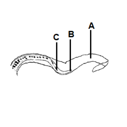
A Sheath
B Tip of tail without nuclei
C Last nucleus