Introduction to Diagnostic Medical Parasitology
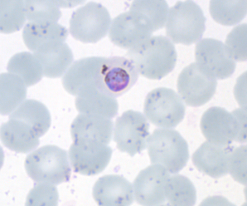
Plasmodium malariae
Growing trophozoite (band form)
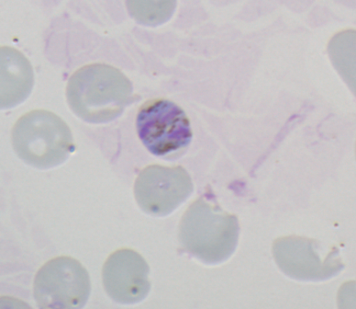

Growing trophozoite (band form)
- Without pseudopodia
- Rounded or bandshaped
- Vacuoles small or absent
- No stippling
- RBCs normal to smaller size
Older trophozoite

Older trophozoite
- Compact cytoplasm
- Less vacuoles
- No stippling
Young schizont

Young schizont
- Enlarged cytoplasm
- four nuclei (chromatin)
- RBC normal to smaller size
- Pigment is beginning to concentrate (brownish/yellowish)
Mature schizont
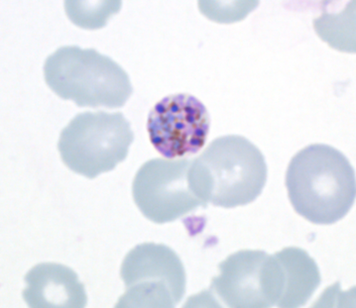
Mature schizont
- Single merozoites visible (up to 8 counts).
Ruptured schizont
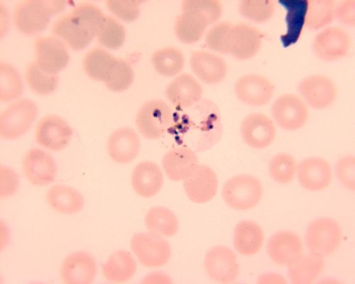
Ruptured schizont
- RBC bursted
- Single merozoites visible (8 counts)
Macrogametocyte
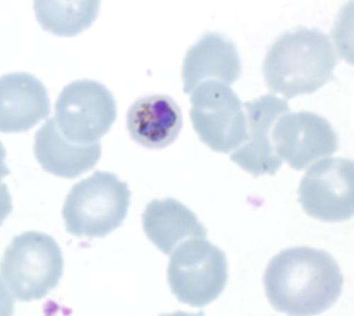
Macrogametocyte
- Chromatin small
- Eccentric and dark red
- Cytoplasm homogeneous
- Blue
- Without vacuoles
- Pigment scattered
Macrogametocyte fills the RBC but smaller than P. vivax