Introduction to Diagnostic Medical Parasitology
Entamoeba coli
Virtual Microscope
2 nuclei cyst
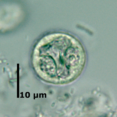
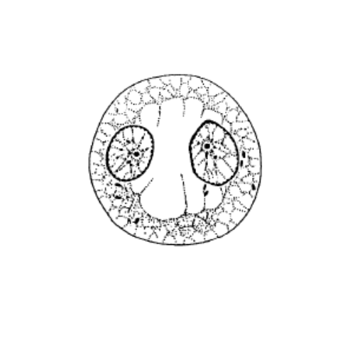
2 nuclei cyst
Size: 15 - 30 μm
- Immature cyst with 2 nuclei
- Typically located on opposite sides
- Only one nuclei in focus
- Most commen stage seen in stool specimen
- 1 and 4 nuclei cysts rarely seen
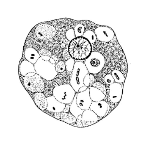
8 nuclei cyst
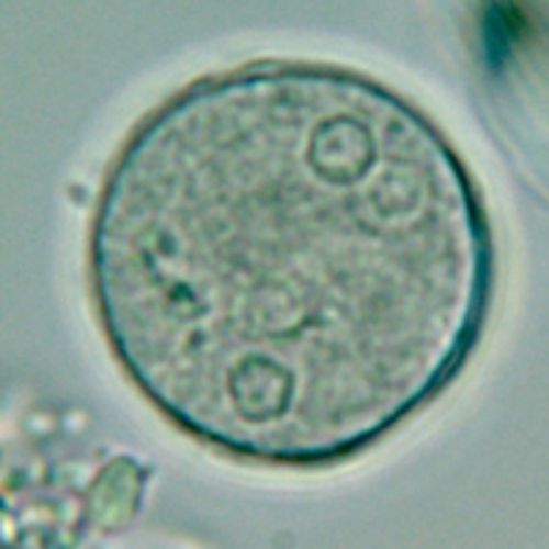
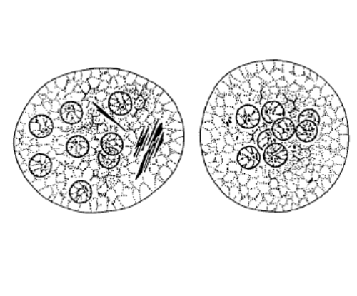
8 nuclei cyst
Size: 15 - 30 μm
- Mature cyst
- Nuclei not always all visible and sometimes centrally gathered
Vegetative form
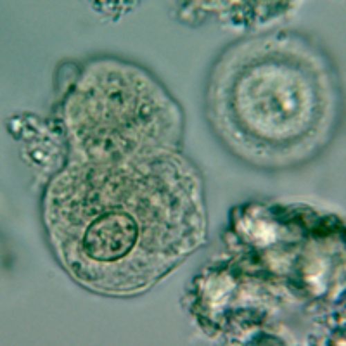
Vegetative form
Size: 15 - 50 μm
- Larger and structures more prominent than in E. histolytica
- Difficult to differentiate from E. histolytica.