Introduction to Diagnostic Medical Parasitology
Chilomastix mesnili
Virtual Microscope
Cyst


Cyst
Size: 7 - 9 x 4 - 6 μm
- Cyst is mostly pear shaped or round
- In the pear shaped form one pole is flattened
- The cyst has a single nucleus
Vegetative form

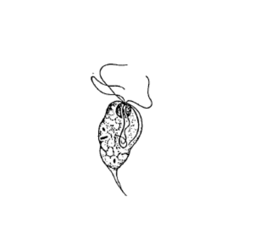
Vegetative form
Size: 15 x 7 μm
- Pear shaped trophozoite with sometimes pointed end and a single nucleus at the opposite end
- Where also the flagella are located