Introduction to Diagnostic Medical Parasitology
Plasmodium falciparum
Rings; accolé form
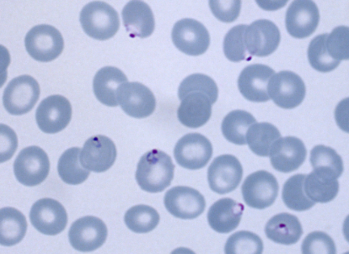
Rings; accolé form
-Delicate rings
- May have two dots of chromatin
- Apliqué or accolé forms (looks like they are attached to erythrocyte membrane)
- All sizes of RBCs
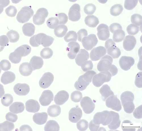
Rings; multiple infection
Rings; multiple infection
- Delicate rings
- May have two dots of chromatin
- Apliqué or accolé forms (looks like they are attached to erythrocyte membrane)
- Multiple infection more frequent
- All sizes of RBCs
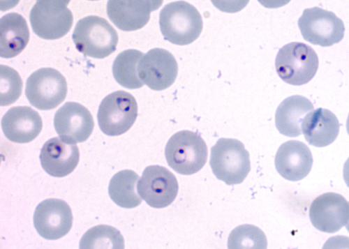
Rings
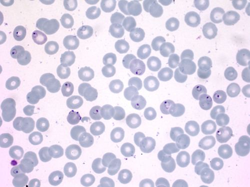
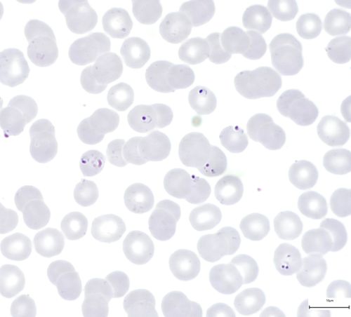
Rings
- Delicate rings
- May have two dots of chromatin
- Apliqué or accolé forms (looks like they are attached to erythrocyte membrane)
- All sizes of RBCs
Older trophozoite with Maurer's dots
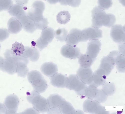
Older trophozoite with Maurer's dots
- In older stages of the trophozoite
- There may appear the Maurer's dots
- They are only visible
- If there was correct pH (7.2) in the staining-solution-buffer
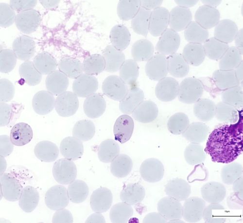
Macrogametocyte
Macrogametocyte
- The gametocytes of P. falciparum have banana form
- The chromatin mass of the macrogametocyte is compact
- Dark red and in the center
-The cytoplasm is blue to violet
- The Pigment is concentrated round chromatin
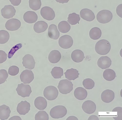
Microgametocyte
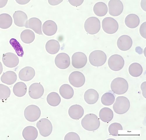
Microgametocyte
- The gametocytes of P. falciparum have banana form
- The form is more stumpy than in the macogametocyte
- Chromatin mass larger
- More diffuse
- Loose
- Light red fragments and filaments
- Cytoplasm from violet to pink
- Always more reddish than in macrogametocyte
- Pigment is scattered.