Introduction to Diagnostic Medical Parasitology
Essentials
Toxocariasis or visceral larva migrans is caused by the migration of nematode larvae to various organs. Human serve as a transport host in which larvae of Toxocara canis (dog as final host) or Toxocara cati (cat as final host) cannot develop to adult worms. Larvae may remain viable in various tissues for years.
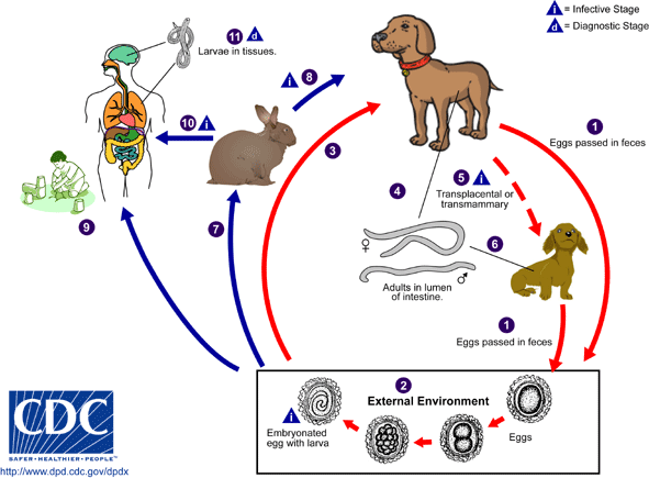
Humans (mainly children) become infected by ingestion of soil contaminated with infective Toxocara eggs in which second-stage larvae have developed. Direct infection from dogs to humans is therefore not possible. Puppies are epidemiologically the most important egg excreters. They become infected by transplacental and transmammary migration of T. canis larvae and pass eggs in their stools by the time they are 3 weeks old. In adult dogs, the infection remains dormant. In humans embryonated eggs hatch in the small intestine, penetrate across the epithelium and are then distributed via the blood in all parts of the body. In rare cases, they may cause disease.

Epidemiology
- Distribution of toxocariasis is worldwide
- Most infections are asymptomatic
- Infections are mainly observed in children aged 14–40 months; adults are less frequently acutely infected
Pathology
- Most infections are without pathological signs
- Pathology is related to worm burden and host responses
- Larvae may damage the eye by inducing inflammatory granulomatous reactions (to be differentiated from retinoblastoma)

Clinical Findings
- Most infections cause no symptoms
- In symptomatic cases, one might observe a variety of signs and symptoms like fever, anorexia, muscle pain, hepatosplenomegaly, lymphadenopathy, abdominal pain, skin reactions or pulmonary symptoms
- In ocular larva migrans, one might observe retinal inflammation, granulomatous reactions, endophthalmitis or uveitis
Diagnosis
Diagnostic methods
Parasitological diagnosis
Clinically suspected diagnosis can only be confirmed by identification of larvae in autopsy or biopsy specimens. Larvae are detected only very rarely (but occasionally in ocular larva migrans). Biopsies not recommended as a routine diagnostic approach
Most infections are without symptoms!
Molecular diagnosis
No test developed so far
Antigen detection
No test developed so far
Antibody detection
Antibody detection by ELISA using excretory/secretory antigens from larval cultures is the standard diagnostic test. Detecting IgG4 antibodies leads to better specificity but with a considerable loss in sensitivity. Serological results cannot differentiate past from active infections. In treated patients, one might observe a decline in antibody titres within 6–12 month post-therapy which might indicate successful treatment.
Diagnostic strategies
Diagnostic approaches are limited to clinical findings and serology!
Prevention and control
- Appropriate disposal of dog faeces
- Protection of children's playgrounds from contamination with dog or cat faeces
- Children should wash their hands after playing with soil or sand
- Systematic deworming of puppies, kittens and lactating female dogs and cats