Introduction to Diagnostic Medical Parasitology
Essentials
Paragonimiasis is caused by lung flukes of the genus Paragonimus. There are several zoonotic species described which infect humans. They all have a complicated life cycle with two intermediate hosts. In addition to humans, dogs and cats can also serve as final hosts.
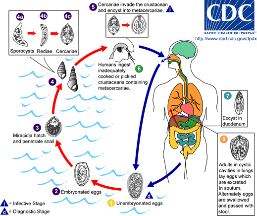
Adult worms live encapsulated in the lungs or in other ectopic locations. Eggs are found in the sputum or when swallowed in the faeces. Within the egg, a miracidium develops and hatches in freshwater. As for all trematodes, the first intermediate host is a snail in which thousands of cercariae develop. These have to find a second intermediate host - crabs and crayfish - in which cercariae encyst in the gills or in the body muscles and develop into metacercariae. Infection in humans results from ingestion of raw infected crabs or crayfish. Metacercariae excyst in the duodenum. Young flukes leave the intestine, migrate within the abdominal cavity, penetrate the diaphragm and invade the pleural cavity.

Epidemiology
- Paragonimiasis is found in tropical and subtropical areas of Asia, Africa, South and Middle America
- Infection sources are uncooked crabs or crayfish
- Domestic cats and dogs can also serve as definitive hosts for Paragonimus spp.


Pathology
- Pulmonary location: mechanic damage and tissue lesions due to toxic metabolites or immunological reactions (eosinophilic inflammatory responses). When cysts burst, eggs are expectorated and bronchopneumonia can result. Cysts can become fibroid and calcified.
- Extrapulmonary localisation is not uncommon. Cysts or abscesses can be found in the liver, brain, subcutis, intestinal wall and other sites
Clinical Findings
- As acute and chronic symptoms in pulmonary infections: fever, chills, cough, haemoptysis, chest pain (X-rays show infiltrate, nodules, cavities, cysts and pleural effusion)
- Worms in the central nervous system can cause meningoencephalitis, seizures, cerebral haemorrhage
- In abdominal paragonimiasis, there are painful abscesses in the liver, spleen or abdominal cavity
- In cutaneous infections, nodules are formed subcutaneously
Diagnosis
Diagnostic methods
Parasitological diagnosis
Ova can be found in sputum or faeces. Humans can be infected with several species (P. westermani, P. heterotremus, P. kellicotti, P. uterobilateralis and others).
WARNING: acid-fast staining for tuberculosis destroys the eggs and precludes diagnosis!
Molecular diagnosis
PCR-RFLP or multiplex PCR allows the species differentiation of metacercariae or adult worms
Antigen detection
No tests developed so far
Antibody detection
Home-made serological tests (mainly ELISAs using crude worm extracts or excretory/secretory antigens) have been developed which all have a problem with specificity. Tests with recombinant antigens have not yet been fully validated.
Diagnostic strategies
Besides parasitological examinations, no special diagnostic strategy can be recommended today!

Prevention and control
Health education: proper handling of food (cooking of crabs and crayfish)