Introduction to Diagnostic Medical Parasitology
Essentials
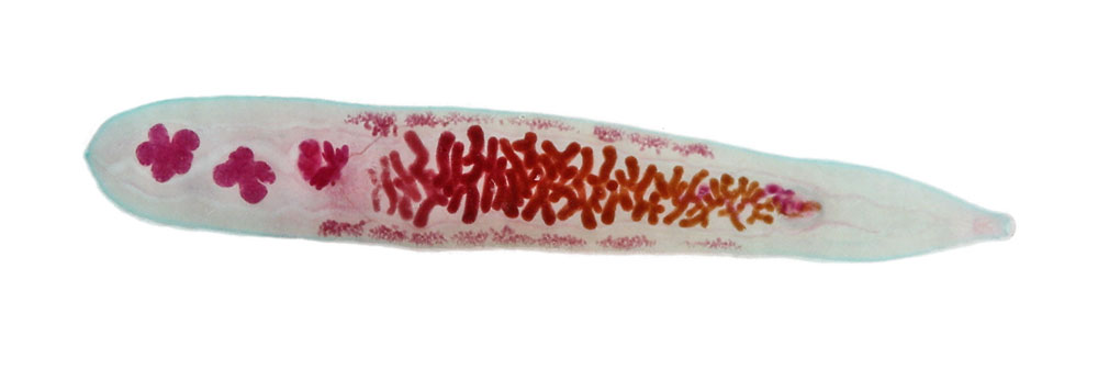
Opisthorchiasis is caused by small liver flukes of cats and other fish-eating animals. The medically most important species are Opisthorchis viverrini and O. felineus. The adult flukes (length up to 1 cm) live in the biliary ducts
The life cycle of Opisthorchis is similar to that of Clonorchis sinensis. Eggs are passed in the faeces of the final host and when miracidia have developed, eggs hatch and motile miracidia infect the first intermediate host, special water snails. In the snail, a further development and multiplication takes place. Cercariae leave the snail and percutaneously infect various freshwater fish which serve as second intermediate hosts. Parasites encyst and form metacercariae. When the fish is eaten by carnivores (humans, dogs, cats), the young flukes emerge and can pass up the common bile ducts, finally reaching the biliary tree, where they mature.
Epidemiology
- Opisthorchis felineus is mainly found in eastern Europe and Russia
- Opisthorchis viverrini is distributed in parts of northern Thailand and Laos
- Infection is caused by eating raw or undercooked fish containing metacercariae
- The worm burden increases with exposure and age
- Opisthorchiasis is a zoonotic infection
Pathology
- Pathology is usually mild and is correlated with the worm load
- In heavy infections, bile ducts may be damaged by inflammation, necrosis, periductal fibrosis and, rarely, calcification
- In severe infections, flukes may enter the liver and cause tissue damage
Clinical Findings
- Many infections are asymptomatic
- Early symptoms are very rare; in chronic infections, symptoms vary: weakness, anorexia, diarrhoea, light fever, liver symptoms and epigastric pain
Diagnosis
Diagnostic methods
Parasitological diagnosis
As for other liver flukes, diagnosis is achieved by detecting eggs in faecal samples. Due to considerable variability in size, it is difficult to distinguish eggs of different Asian liver fluke species.
Molecular diagnosis
There have been first attempts to detect and differentiate DNA from Asian liver flukes (C. sinensis, O. viverrini and O. felineus) in faecal samples using specific primers
Antigen detection
No tests developed so far
Antibody detection
Serological tests using crude worm antigens have good sensitivity but low specificities (cross-reactions with other trematode infections).
A better specificity – with a considerable loss in sensitivity! – can be achieved using recombinant or purified antigens (e.g. cysteine proteinase or excretory/secretory antigens).
Diagnostic strategies
- To diagnose a symptomatic case
Diagnosis is achieved by detecting ova in one or two stool samples
- To screen an exposed population
One might use a serological method if only one trematode fluke species is prevalent in the study area

Prevention and control
- In endemic areas, people should be educated about the dangers of eating raw or undercooked fish (by freezing fish, metacercariae are killed!)
- Faeces should be disposed of in a sanitary way
- No night soil or animal waste should be deposited in fish ponds
