Introduction to Diagnostic Medical Parasitology
Essentials
The small tapeworm (length under 1 cm) of dogs and canids, Echinococcus granulosus, is the most common species of Echinococcus. Intermediate hosts include herbivores, sheep, cattle, pigs, horses and omnivore animals. After an accidental infection, humans man can also become an intermediate host, resulting in cystic hydatid disease.
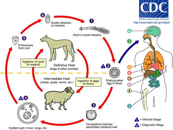
Dogs and canids are definitive hosts for Echinococcus granulosus. Eggs are passed with the faeces. After per oral uptake of eggs by an intermediate host (e.g. sheep or cattle), the oncosphere emerges from the egg and invades tissues of the intermediate host, primarily the liver. There the development of hydatid cysts (a fluid-filled larval stage with an inner wall with multiple scolices) takes place. If the infected intermediate host is eaten by a carnivore, scolices evert in the intestine and grow to adult tapeworms.


Epidemiology
- Distribution is worldwide (mainly in temperate zones)
- The zoonotic infection is caused by hand-to-mouth transfer of tapeworm eggs from infected dogs, or indirectly by contaminated food, water or soil
- Hydatid disease (in most cases only a single cyst) becomes clinically manifest only after many years

Pathology
- Hydatid cysts are mainly found in the liver (65%), the lungs (25%) and other organs (e.g. brain) and are for many years well tolerated
- Symptoms start when cysts reach a certain size (over 10 cm)
- Rupture of cysts might provoke anaphylactic reactions

Clinical Findings
- Many patients are asymptomatic
- An intact hepatic cyst might provoke the following signs and symptoms: liver enlargement, right epigastric pain, nausea, vomiting
- The rupture of a liver cyst might lead to acute allergic reactions (anaphylaxis), cholangitis, jaundice, portal hypertension, and eventually to a hepatic abscess by secondary infections
- After rupture, a lung cyst may cause chest pain, cough, dyspnoea and haemoptysis
- A brain cyst might cause epilepsy


Diagnosis
Diagnostic methods
Parasitological diagnosis
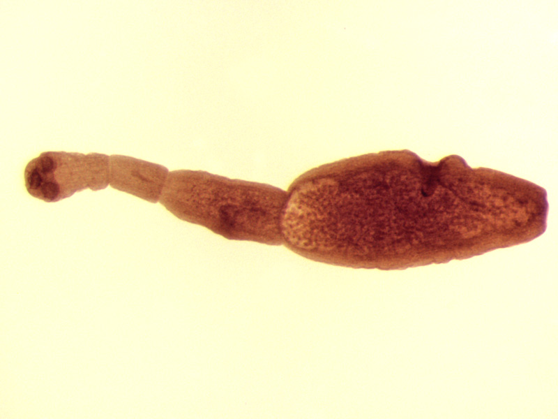
Parasitological diagnosis is only possible by detecting scolices, daughter cysts or hooklets (“hydatid sand”) in the sediment of the hydatid fluid.
Clinical diagnosis is based on signs and symptoms of slow-growing tumours and history of residence in endemic areas, along with association with canines (e.g. in connection with slaughterhouses).
Radiology, CT and sonography along with serologic tests are useful for the diagnosis of hydatid disease caused by Echinococcus granulosus.
Molecular diagnosis
PCR with excellent sensitivity and specificity is used to detect parasite nucleic acids in needle aspirates or to detect copro-antigens in dogs.
Antigen detection
Several home-made tests detect copro-antigens in the faeces of definitive hosts by ELISA and specific monoclonal antibodies. One test kit is commercially available.
Antibody detection
Serology is an important diagnostic element for detecting hydatid disease. Various test formats (IFA, ELISAs, Western blots) have been developed to detect specific antibodies.
Using an affinity-purified or recombinant antigen (e.g. Em18) a differential diagnosis between the two species seems possible.
Diagnostic strategies
- To diagnose an individual case
Combining imaging techniques with serology is the best strategy today
- To assess endemicity in an exposed population
Serology is the first choice!

Prevention and control
- Personal hygiene must be emphasised to prevent accidental ingestion of the cestode eggs from soil contaminated with dog faeces or by direct transmission
- All infected viscera from slaughtered animals must be disposed of so that dogs do not have access to this material
- Periodic anti-helminthic treatment of dogs in endemic areas