Introduction to Diagnostic Medical Parasitology
Essentials
Dracunculiasis or guinea or Medina worm infection is caused by the tissue nematode Dracunculus medinensis. This worm has an indirect life cycle with small copepods as intermediate hosts. Adult worms live in the connective and subcutaneous tissues, particularly in the legs. Dracunculiasis has a high morbidity and causes economic loss by reducing the working capacities of affected individuals. Worldwide attempts to eradicate this disease have met with initial success: the number of infected people has decreased significantly!
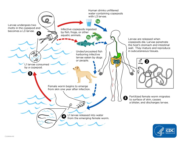
Humans get infected by drinking water containing water fleas or copepods (Cyclops spp.) infected with third-stage larvae (approx. 0.5 mm in length). Infective larvae penetrate the intestinal wall and migrate via lymphatics to connective tissues where they develop into adult worms.

Epidemiology
- Guinea worm infection is endemic on the Indian subcontinent, in the Middle East, and in central and western Africa, mainly in rural areas
- Control has been achieved through international efforts in many countries except in areas with social unrest. However, eradication remains a distant goal!
- Infection occurs by ingestion of infected copepods with the drinking water
Pathology
- Blister formation induced by toxic products of the worm
- Death and disintegration of adult worms produce bacteriologically sterile abscesses
Clinical Findings
- Incubation period is about 1 year
- No symptoms during the development of worms
- Skin manifestations (urticaria, erythaema, pruritus) and eventually systemic allergic symptoms before blisters erupt
- Blisters are painful and burning. People become invalided. After a few days the blisters rupture and the anterior end of the female worm is visible. The worm (up to 1.2 m in length!) can be removed slowly.


Diagnosis
Diagnostic methods
Parasitological diagnosis
Dracunculiasis is diagnosed clinically by the effect of the gravid female worm (blister, then skin ulcer) or by an abscess induced by a dying worm). The adult worm may be seen protruding from the skin lesion.
Calcified worms may be seen by X-ray.
Molecular diagnosis
No tests developed
Antigen detection
Attempts to demonstrate D. medinensis antigens have so far been unsuccessful
Antibody detection
Immunodiagnostic tests have been explored using adult female worm and first-stage larval antigens. The most promising first results were obtained by detecting specific IgG4 antibodies.
Diagnostic strategies
Clinical findings are characteristic and no special diagnostic strategy is needed.
Prevention and control
- The easiest personal control measure is to filtrate water before use
- Health education
- To improve the water supply of affected communities
