Introduction to Diagnostic Medical Parasitology
Essentials
Cysticercosis is caused by the larval stage of Taenia solium. Humans can be an accidental intermediate host. Eggs or proglottids of the pork worm, once swallowed, hatch in the small intestine from where the larvae migrate to subcutaneous tissues, striated muscles and other tissues and vital organs (e.g. the central nervous system), where they form cysticerci. Neurocysticercosis is a severe disease.
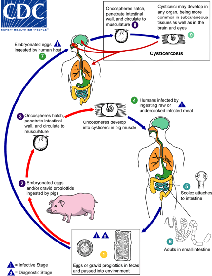
Infections occur after per oral uptake of embryonated eggs with faecally contaminated uncooked food or soil. Eggs are only infective when they contain a second-stage larva. Therefore no direct person-to-person transmission is possible. This development outside the host takes 2 to 3 weeks and the eggs remain infective for years under optimal conditions. After ingestion of embryonated eggs, larvae hatch in the duodenum and penetrate the mucosa. Via lymphatics and blood, after 2 weeks they reach the lung where they moult and invade alveoli. Larvae ascend the trachea and are swallowed. Adult worms are found in the jejunum approx. 2 months post-infection. Female worms (20 to 40 cm long) produce an average of 200,000 eggs per day!


Epidemiology
- Distribution is worldwide (prevalence is high in parts of Latin America, Africa and South-East Asia where pigs and humans live in close contact)
- Humans as the only definitive host of the tapeworm become intermediate hosts by swallowing T. solium eggs with contaminated food or water
- Flies and other insects can serve as mechanical vectors
- Autoinfection is possible
Pathology
- Many cases are without symptoms even if the brain is involved
- Pathology might be provoked by dying cysticerci which induce inflammatory reactions
- Localisation of cysticerci in the CNS and heart may lead in rare cases to fatal disease

Clinical Findings
- In neurocysticercosis one might find the following signs and symptoms: epileptiform seizures, intracranial hypertension with nausea and vomiting, neurologic psychiatric disturbances
- Special localisations of cysticerci can cause a variety of syndromes


Diagnosis
Diagnostic methods
Parasitological diagnosis
Cysticerci (tapeworm larvae) of T. solium occur in the muscle and in the brain (in humans as intermediate hosts).
Subcutaneous cysticerci may be visible or palpable.
Cysticerci may also be detected by non-invasive methods like CT scan and MRI, or when calcified, by X-ray.
Histology reveals cysticerci in biopsies.
Molecular diagnosis
No tests developed so far
Antigen detection
First attempts to detect antigens in cerebrospinal fluid have been described
Antibody detection
Diagnosis of cysticercosis should be supported by a serological test. Serum antibody detection in cysticercosis is done with immunoassays (enzyme-linked immunotransfer blots, EITB). No advantage was observed using cerebrospinal fluid.
Diagnostic strategies
- To diagnose a symptomatic patient
For neurocysticercosis, imaging techniques should be combined with serology. Subcutaneous cysticerci may be visible or palpable, or when already calcified, detectable by X-ray.
- To screen an exposed population
Serology is the method of choice

Prevention and control
- Educate people to prevent faecal contamination of soil, water, human and animal food by personal hygiene and sanitation
- Measures that prevent humans becoming a final host (see under Taeniasis)
- Mass treatment of infected humans to prevent transmission in endemic areas